Laryngeal

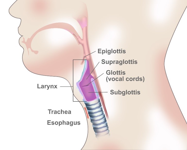
Laryngeal cancer is a disease in which malignant (cancer) cells form in the tissues of the larynx–the part of the throat between the base of the tongue and the trachea. The larynx contains the vocal cords, which vibrate and make sound when air is directed against them. The sound echoes through the pharynx, mouth, and nose to make a person’s voice.

There are three main parts of the larynx, which include:

  • Supraglottis – the upper part of the larynx above the vocal cords, including the epiglottis, which is the flap that covers the trachea when swallowing so that food does not enter the lungs.
  • Glottis – the middle part of the larynx where the vocal cords are located.
  • Subglottis – the lower part of the larynx between the vocal cords and the trachea (windpipe).

Laryngeal cancer is a type of head and neck cancer that forms in squamous cells, which are the thin, flat cells that line the inside of the larynx.

Anything that increases your risk of getting a disease is called a risk factor. Risk factors of laryngeal cancer may include:

  • Smoking and/or chewing tobacco
  • Moderate or heavy alcohol use (more than one drink a day)
  • Poor nutrition
  • Human papillomavirus (HPV) infection
  • Genetic syndromes (inherited gene mutations)
  • Workplace exposures to certain fumes and chemicals
  • Gender (more common in men than women)
  • Age (more common among patients age 65 and older)
  • Race (more common among African Americans and Caucasians)
  • Gastroesophageal reflux disease (GERD)

Keep in mind that having a risk factor, or even several of them, does not mean that you will get laryngeal cancer. Likewise, many people who get the disease may have few or no known risk factors.

There are several signs and symptoms that could be related to laryngeal cancer; however, it is important to remember that they can also be symptoms for other diseases. See your doctor if you have:

  • A change in your voice, such as unexplained hoarseness
  • A lump in the neck
  • Persistent sore throat
  • Ear pain not caused by an ear infection or other condition
  • Pain or difficulty when swallowing

Again, these could be symptomatic of something other than cancer. With that said, if any of these symptoms last for more than three weeks, it is a good idea to be checked by a doctor. If it is cancer, early detection can give you better treatment results.

There are a variety of tests that examine the throat and neck in order to detect and diagnose laryngeal cancer. In many cases you will start with an ear, nose, and throat specialist (ENT) who will perform the exams and request further testing if your doctor believes there may be a tumor in the larynx.

If the doctor suspects there could be cancer, a biopsy may be performed.  A biopsy requires a tissue sample be collected from the area of the body where cancer is suspected so that the cells can be tested to see if cancer is present. Some of the following tests or procedures may be run to make a determination.

  • Physical exam of the throat – An exam in which the doctor feels for swollen lymph nodes in the neck and looks down the throat with a small, long-handled mirror to check for abnormal areas.
  • Laryngoscopy – A procedure to look at the larynx (voice box) for abnormal areas. A mirror or a laryngoscope (a thin, tube-like instrument with a light and a lens for viewing) is inserted through the mouth to see the larynx. A special tool on the laryngoscope may be used to remove samples of tissue.
  • Endoscopy – A procedure used to look at areas in the throat that cannot be seen with a mirror during the physical exam of the throat. An endoscope (a thin, lighted tube) is inserted through the nose or mouth to check the throat for anything that seems unusual. Tissue samples may be taken for biopsy.
  • Panendoscopy – A procedure that combines laryngoscopy, esophagoscopy, and (at times) bronchoscopy. This lets the doctor thoroughly examine the entire area around the larynx and hypopharynx, including the esophagus (swallowing tube) and trachea (windpipe). While the patient is under general anesthesia, the doctor will thoroughly examine all of these areas to look for tumors and determine their size and if they’ve spread. A tissue sample can be collected for biopsy during this procedure as well.

If cancer has been found in the biopsy, it’s common to add other tests including imaging to see if the cancer has spread, and if so, how far. Your doctor may request one or more of the following imaging studies:

  • CT scan (CAT scan) – A procedure that makes a series of detailed pictures of areas inside the body, taken from different angles. The pictures are made by a computer linked to an X-ray machine. A dye may be injected into a vein or swallowed to help the organs or tissues show up more clearly. This procedure is also called computed tomography, computerized tomography, or computerized axial tomography.
  • PET scan (positron emission tomography scan) – A procedure to find malignant tumor cells in the body. A small amount of radioactive glucose (sugar) is injected into a vein. The PET scanner rotates around the body and makes a picture of where glucose is being used in the body. Malignant tumor cells show up brighter in the picture because they are more active and take up more glucose than normal cells do. In some cases the PET scan and CT scan may be used together. This is called a PET-CT.
  • MRI (magnetic resonance imaging) – A procedure that uses a magnet, radio waves, and a computer to make a series of detailed pictures of areas inside the body. This procedure is also called nuclear magnetic resonance imaging (NMRI).
  • Bone scan – A procedure to check if there are rapidly dividing cells, such as cancer cells, in the bone. A very small amount of radioactive material is injected into a vein and travels through the bloodstream. The radioactive material collects in the bones with cancer and is detected by a scanner.
  • Barium esophagogra – An x-ray of the esophagus that is taken after the patient drinks a liquid that contains barium (a silver-white metallic compound). The liquid coats the esophagus and makes it easier for the doctor to see problems in the throat.

Many of the test results (as described in the Diagnosis section) are used to determine the extent, or stage, of the cancer. The stage of cancer describes how much cancer is in the body. It helps your doctor understand the seriousness of the cancer, how best to treat it, and what the chance of survival is.

For laryngeal cancer, doctors mostly depend on the TNM system created by the American Joint Committee on Cancer (AJCC). The TNM system is based on three key pieces of information:

  • How big the main tumor (T) is
  • If the cancer has spread to nearby lymph nodes (N)
  • The spread (metastasis) to distant parts of the body (M)

Numbers or letters after T, N, and M provide more details about each of these factors. Lower numbers mean that the cancer is in an early stage. Higher numbers mean the cancer is more advanced.

Hypopharyngeal

Hypopharyngeal cancer is a disease in which malignant (cancer) cells form in the tissues of the hypopharynx. It is a type of head and neck cancer. 

The hypopharynx is the bottom part of the pharynx (throat). The pharynx is a hollow tube about five inches long that starts behind the nose, goes down the neck, and ends at the top of the trachea (windpipe) and esophagus (the tube that goes from the throat to the stomach ). Air and food pass through the pharynx on the way to the trachea or the esophagus. 

Most hypopharyngeal cancers form in squamous cells, the thin, flat cells lining the inside of the hypopharynx. The hypopharynx has three different areas. Cancer may be found in one or more of these areas.